Lisa Lobel and Ben McFarland
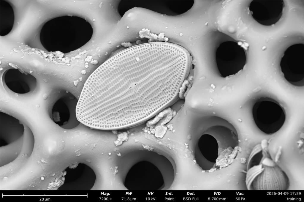
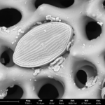
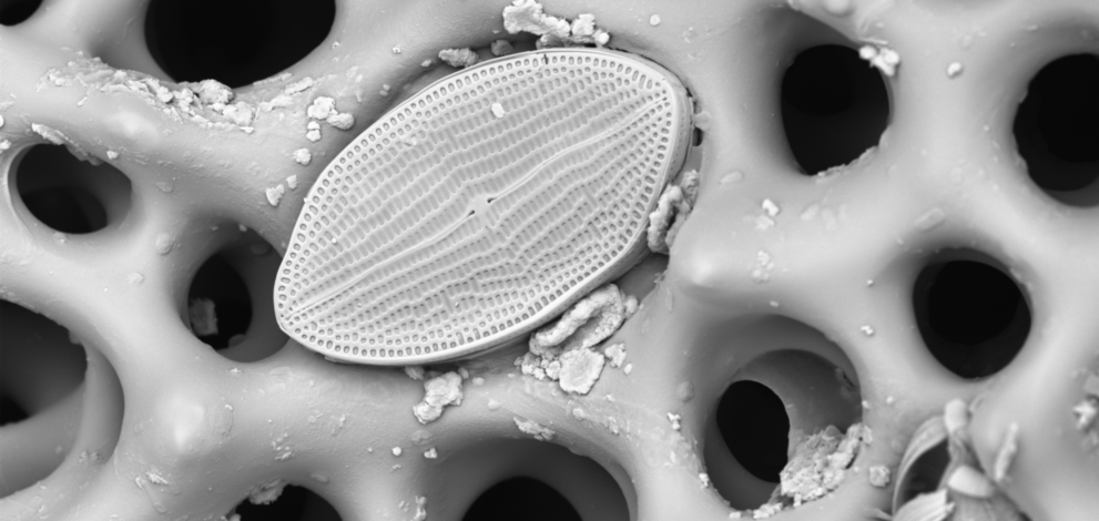
The sea urchin samples were collected by Professor Lobel on the Mesoamerican Barrier Reef in Belize. Photomicrograph taken with a Phenom (Thermo Scientific) desktop Scanning Electron Microscope at 7200X. The sample of sea urchin endoskeleton (test) was first prepared by breaking a section to fit onto the SEM sample stage and then sputter coated with gold for 40 seconds using a Cressington Sputter coater 108. The sample was scanned for interesting features including the diatoms that were stuck in the various skeletal pores. Diatoms are unicellular phytoplankton, and this diatom is likely to be in the genus Mastoglia. It is approximately 20 x 30 microns in size.